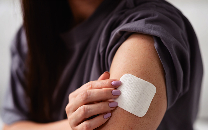
In 2020, Scientific American recognized microneedle technology, known for its painless injections and tests, as one of the “Top 10 Emerging Technologies that Could Change the World.” Compared to traditional drug delivery methods, microneedling presents several advantages, including bypassing the first-pass effect, minimizing pain, enhancing patient compliance, and allowing controlled release, all of which enhance its market potential. This article provides an overview of microneedling, pharmacokinetic studies in large animals, and our suggestions for addressing its challenges.
Overview of microneedle
Microneedles consist of an array of micron-sized needle tips attached to a base. Their lengths range from 100 to 3000 μm, with diameters of 50 to 250 μm, typically using lengths between 250 and 1500 μm, depending on their specific applications [1]. The concept of transdermal drug delivery using microneedles was first introduced by Gerstel and Place in 1971[2]. In 1995, Hasmahi et al. [3] successfully fabricated microneedles using silicon materials. It wasn’t until 1998 that Mark Prausnitz [4] and his team applied microneedles for transdermal drug delivery, validated their feasibility, and published their findings. In recent years, microneedle patches have been utilized for skin administration as medical devices, drawing regulatory attention from authorities.
Mechanism and advantages of microneedling
Microneedling is an innovative drug delivery method that uses micron-sized needles to penetrate the skin’s stratum corneum, creating temporary microchannels to release drugs directly into the epidermis or dermis. The drug then enters the systemic circulation through capillaries, enabling systemic treatment. Because microneedles are typically only a few hundred micrometers in length, they are sufficient to insert into the skin without reaching pain nerves, allowing for painless or minimally painful drug delivery.

Figure 1. Mechanism of drug delivery by microneedle device [5]
Microneedling offers a range of advantages over traditional transdermal delivery systems, such as bypassing the hepatic first-pass effect and the degradation of the drug in the gastrointestinal tract, maintaining stable blood drug concentrations, ease of use, and controlled or sustained release. Additionally, microneedle technology addresses the limitations of traditional transdermal systems, such as the challenges of delivering biomacromolecules and hydrophilic compounds through the skin and the associated low bioavailability. Compared to the commonly used methods for macromolecular drug delivery intravenous or subcutaneous injections, microneedles enable minimally invasive drug delivery, reducing or eliminating pain, significantly improving patient compliance. Moreover, after the microneedles dissolve or are removed, the microchannels they create in the skin close, restoring the barrier function and preventing skin infections [6].
Overview of microneedle types and applications
Microneedles can be broadly classified into five categories based on drug delivery strategies: solid, coated, hollow, dissolvable, and hydrogel-forming microneedles [7].
Solid microneedles: Solid microneedles are usually made from silicon, metal, or non-degradable polymer materials. Their high mechanical strength allows them to effectively insert into the skin to create microchannels. Consequently, drug delivery with solid microneedles requires two steps: (1) Puncturing the skin to create microchannels; and (2) applying the drug to the microchannel area.
As the earliest type of microneedle developed, solid microneedles also have clear disadvantages. This two-step process is less convenient and lacks precise dose control. Currently, solid microneedles are primarily used in the aesthetic medicine industry.
Coated microneedles: To avoid the two-step drug delivery process, researchers have developed coated microneedles based on solid microneedles. Drugs are applied to the microneedles’ surface through immersion, spraying, or piezoelectric inkjet printing. Upon penetrating the skin, the coating dissolves, releasing the drug. However, the limited surface area of microneedles restricts their drug-loading capacity.
Hollow microneedles: Hollow microneedles are similar to micron-sized syringes/needles, featuring a needle structure with an internal lumen that can penetrate the stratum corneum and deliver drugs through its internal channel. While hollow microneedles are also commonly made from silicon or metal materials, their unique internal structure makes them prone to breakage compared to solid microneedles.
Dissolvable microneedles: Dissolvable microneedles are one of the most popular types of microneedles currently being developed. They are primarily fabricated from soluble or biodegradable polymers or carbohydrates with good biocompatibility, low toxicity, and high plasticity [8]. The fabrication process for dissolvable microneedles is relatively simple and mild, making them more beneficial to protein and peptide drugs, allowing them to maintain long-lasting biological activity [7]. Dissolvable microneedles can be used for the delivery of biomacromolecule drugs, such as vaccines, antibodies, hormones, and nucleic acid drugs [9].
Hydrogel-forming microneedles: Hydrogel-forming microneedles are also one of the popular types of microneedles, typically made from crosslinked polymer materials. They can penetrate the stratum corneum and quickly absorb interstitial fluid, causing the microneedles to expand and release the drug. After expansion, hydrogel-forming microneedles can resist the closure of skin microchannels to a certain extent, enabling higher drug loading capacity and sustained drug release. Additionally, the drug release rate can be controlled by selecting the crosslinking density of the materials used in fabrication.
As technology advances and personalized needs grow, microneedle types continue to innovate, including cryo-microneedles, separable microneedles, and irregularly shaped designs [8].
Applications of microneedles
In recent years, major research institutions around the world have been actively developing microneedle formulations. Microneedling is being widely applied in drug delivery, vaccination, biosensing, disease diagnosis, and aesthetics. The table below lists microneedle projects registered with the NIH and NMPA.
|
Study Title |
Registration number |
Intervention / Treatment |
Conditions |
|
Dexmedetomidine hydrochloride microneedle patch |
CXHL2400156 CXHL2400157 |
Dissolvable microneedles |
Pediatric preoperative sedation |
|
Double-Blind Comparison of the Efficacy and Safety of C213 to Placebo for the Acute Treatment of Cluster Headaches |
NCT04066023 |
The zolmitriptan-coated titanium microneedle array |
Cluster headache |
|
Insulin Delivery Using Microneedles in Type 1 Diabetes |
NCT00837512 |
Microneedles, subcutaneous insulin catheters |
Type 1 Diabetes Mellitus |
|
Efficacy of Transdermal Microneedle Patch for Topical Anesthesia Enhancement in Paediatric Thalassemia Patients |
NCT05078463 |
Microneedles; Eutectic Mixture of Local Anesthetics (EMLA) Cream |
Anesthesia |
|
Measles and Rubella Vaccine Microneedle Patch Phase 1-2 Age De-escalation Trial |
NCT04394689 |
Measles-Rubella Vaccine microneedle patch |
Measles, rubella |
|
Microneedle Array Plus Doxorubicin in Cutaneous Squamous Cell Cancer (cSCC) |
NCT05377905 |
Microneedle Array Doxorubicin |
Cutaneous Squamous Cell Carcinoma |
|
Inactivated Influenza Vaccine Delivered by Microneedle Patch or by Hypodermic Needle |
NCT02438423 |
Microneedles; Inactivated Influenza Vaccine |
Influenza |
Table 1. Microneedle projects registered with NIH and NMPA [8]
In vivo pharmacokinetic studies of microneedles
In vivo absorption of transdermal drugs is crucial for evaluating the quality of transdermal formulations. Pig skin, which closely resembles human skin in histological and biological structures, makes laboratory pigs an ideal model for transdermal evaluation studies. Performing in vivo pharmacokinetic studies with laboratory pigs allows for a better understanding of the absorption profiles of microneedle formulations, offering valuable insights for research and optimization in microneedle product development.
Case study of microneedle application
This study evaluated the skin insertion of dissolvable liraglutide microneedles based on a minipig model (Figure 2). Following insertion, the microneedle tips dissolved within the epidermis (Figure 2a), leaving clear gentian violet-stained pinholes in the skin (Figure 2b).

Figure 2. The morphology of dissolvable liraglutide microneedles before (a-1) and after (a-2) insertion into pig skin;
(b) Pig skin stained with 1% gentian violet solution after insertion [10]
In this study, researchers administered minipigs with liraglutide by dissolvable microneedles or SC injection once daily for 7 days. It was clearly shown that both groups reached a steady state after three consecutive administrations, as illustrated in Figure 3a. The Tmax of dissolvable microneedles after the first and last administrations were significantly shorter than that of SC injection (Figure 3a & Figure 3b), indicating that transdermal drug absorption following dissolvable microneedles was faster than that of SC injection.

Figure 3. Plasma drug concentration-time profiles of liraglutide after administration to minipigs [10]
After evaluating skin irritation from microneedling, it was found that no skin irritation was observed in minipigs except for mild erythema occurring within 4 h after once-daily administration for 7 days at the same (Figure 4), indicating that dissolvable liraglutide microneedles were well tolerated.

Figure 4. Skin irritation after application of dissolvable liraglutide microneedles to minipigs [10]
These above results suggest that dissolvable microneedles might offer a safe and effective alternative to SC injection for the administration of liraglutide.
Our experience in microneedle projects
Administering microneedles to minipigs presents several challenges:
-
Unlike traditional oral or intravenous injections using syringes, microneedle patches are small, making them difficult to handle and manipulate;
-
Different types of microneedles have varying mechanical strengths, and improper handling can lead to bending or breaking of the microneedles during administration;
-
The Bama miniature pigs are naturally active and tend to root and rub, complicating the administration process.
To overcome these challenges, WuXi AppTec DMPK has developed various drug administration tools independently, improved restraint slings, and optimized manipulation techniques. These efforts have significantly increased the success rate of microneedling. While administering the same drug to minipigs via dissolvable microneedle and SC injection, the results indicated faster drug absorption through microneedle administration, consistent with findings reported in the literature [10].
Selection of administration site
In addition to optimizing the microneedle manipulation techniques, the selection of an administration site is equally important. The thickness of the skin at the administration site significantly impacts microneedling, directly affecting the success, microneedle penetration depth, and drug delivery efficiency. We have assessed the skin thickness at different sites on minipigs and found that the variation in skin thickness aligns with findings reported in the literature (Fig. 5) [11].

Figure 5. The skin thickness of different sites [11]
Through long-term exploratory testing of the skin at different sites for microneedling on minipigs aged 1 to 5 months, we have summarized the advantages and disadvantages of different skin sites for microneedle administration. The details are shown in Table 2.
|
Administration site |
Advantages |
Disadvantages |
|
Shoulder and back |
|
|
|
Abdomen |
|
|
|
Leg |
|
|
|
Ear |
|
|
Table 2. Comparison of different skin sites for microneedling
Based on the comparison of advantages and disadvantages, ear skin is more conducive to microneedling and enhances drug absorption. Therefore, we selected ear skin as the administration site and developed a comprehensive protocol for microneedle administration.
WuXi AppTec DMPK team has completed dozens of microneedle projects using minipigs and has established a comprehensive protocol for microneedling and patch retrieval. Through these projects, the team has accumulated significant experience and refined methodologies tailored to projects.
A final word
Microneedling represents an innovative direction in modern drug delivery, with continuous advancements driving progress in the field. It brings new solutions to areas such as drug delivery, disease diagnosis, biosensing, vaccine administration, and aesthetic medicine. WuXi AppTec DMPK has extensive experience in in vivo pharmacokinetic studies. Our teams have completed dozens of microneedle projects on minipigs, selected suitable skin sites for microneedling, and established a comprehensive microneedle experimental procedure. These efforts support the development of microneedle formulations and advance progress.
Authors: Qian Jiang, Chen Ning, Huan He, Jiarui Yu, Zhihai Li, Shoutao Liu
Talk to a WuXi AppTec expert today to get the support you need to achieve your drug development goals.
Committed to accelerating drug discovery and development, we offer a full range of discovery screening, preclinical development, clinical drug metabolism, and pharmacokinetic (DMPK) platforms and services. With research facilities in the United States (New Jersey) and China (Shanghai, Suzhou, Nanjing, and Nantong), 1,000+ scientists, and over fifteen years of experience in Investigational New Drug (IND) application, our DMPK team at WuXi AppTec are serving 1,600+ global clients, and have successfully supported 1,500+ IND applications.
Reference
[1] Pettis R J, Harvey A J. Microneedle delivery: clinical studies and emerging medical applications[J]. Therapeutic delivery, 2012, 3(3): 357-371.
[2] Gerstel M S, & Place V A. Drug delivery device. US Patent 3964482-A. 1971.
[3] Hashmi S, Ling P, Hashmi G, et al. Genetic transformation of nematodes using arrays of micromechanical piercing structures[J]. BioTechniques, 1995, 19(5): 766-770.
[4] Henry S, McAllister D V, Allen M G, et al. Microfabricated microneedles: a novel approach to transdermal drug delivery[J]. Journal of pharmaceutical sciences, 1998, 87(8): 922-925.
[5] Waghule T, Singhvi G, Dubey S K, et al. Microneedles: A smart approach and increasing potential for transdermal drug delivery system[J]. Biomedicine & pharmacotherapy, 2019, 109: 1249-1258.
[6] Huang Y, Ma F, Zhan H, et al. Microneedle array used for transdermal delivery of biomacromolecules[J]. Progress in Biochemistry and Biophysics, 2017, 44(9): 757-768
[7] Liu T, Chen M, Fu J, et al. Recent advances in microneedles-mediated transdermal delivery of protein and peptide drugs[J]. Acta Pharmaceutica Sinica B, 2021, 11(8): 2326-2343.
[8] Zhao H, Chen M, Lu A, et al. The application of microneedles in transdermal drug delivery systems [J]. Progress in Pharmaceutical Sciences, 2024, 48(4): 244-253
[9] Wu W, Zhou C, Wu C, et al. Advances in microneedle-mediated transdermal delivery of biomacromolecular drugs[J]. Progress in Pharmaceutical Sciences, 2024, 48(4): 254-268
[10] Lin H, Liu J, Hou Y, et al. Microneedle patch with pure drug tips for delivery of liraglutide: pharmacokinetics in rats and minipigs[J]. Drug Delivery and Translational Research, 2024: 1-15.
[11] Zou Q, Yuan R, Zhang Y, et al. A single-cell transcriptome atlas of pig skin characterizes anatomical positional heterogeneity[J]. Elife, 2023, 12: e86504.
[12] Simon G A, Maibach H I. The pig as an experimental animal model of percutaneous permeation in man: qualitative and quantitative observations–an overview[J]. Skin Pharmacology and Physiology, 2000, 13(5): 229-234.
[13] Jacobi U, Kaiser M, Toll R, et al. Porcine ear skin: an in vitro model for human skin[J]. Skin Research and Technology, 2007, 13(1): 19-24.
Stay Connected
Keep up with the latest news and insights.


